Ⅰ Principle of Super Large Depth of Field Digital Microscope
The principle of Super Large Depth of Field Digital Microscopes primarily involves technologies such as optical imaging, depth-of-field extension, and image fusion. The specifics are as follows:
Optical Imaging Principle: Similar to traditional optical microscopes, Super Large Depth of Field Digital Microscopes form an image of the object on the focal plane of the eyepiece via the objective lens, which is then magnified by the eyepiece for observer viewing. They utilize the refraction principle of lenses to focus light from the object and form a magnified real image, which is further magnified by the eyepiece, allowing the observer to see the details of the object.
Depth-of-Field Extension Technology:
Continuous Zoom System: Super Large Depth of Field Digital Microscopes are typically equipped with continuous zoom objective lenses, allowing the magnification to be changed continuously within a certain range. By observing and capturing images at different magnification levels and utilizing the differences in depth of field at these various magnifications, more clear information about the object's surface can be acquired.
Scanning Technology: Employs a scanning method for object imaging. For example, by moving the sample stage or the objective lens, the object is scanned and captured at different height positions. This allows for acquiring a series of images at different focal planes, each containing clear information from different parts of the object, thereby extending the overall depth of field range.
Image Fusion Principle: Multiple images acquired at different focal planes or magnifications are fused. Through specialized image fusion algorithms, the in-focus parts of these images are extracted and merged into a single image with a large depth of field. Common algorithms include feature-based fusion algorithms, pyramid-based fusion algorithms, etc. These algorithms can automatically identify feature points or regions in the images, match and fuse them, ensuring the final image has sharp details across the entire object surface, presenting a deep-depth-of-field effect.
Ⅱ What Can a Super Large Depth of Field Digital Microscope Measure?
Super Large Depth of Field Digital Microscope can observe and measure various microscopic and macroscopic objects, mainly including the following aspects:
1. Object Dimension Measurement
Length Measurement: Precisely measures the linear length of objects, such as the diameter of metal wires or the side length of tiny components.
Width Measurement: Accurately measures the width of objects of various shapes, such as the width of sheet materials or the line width in integrated circuits.
Height Measurement: Measures the height or thickness of objects by scanning different focal planes, such as the height of electronic components on a circuit board or the thickness of a coating.
Angle Measurement: Measures angles on objects, such as the chamfer angle of mechanical parts or the interplanar angle of crystals.
2. Object Surface Topography Observation and Measurement
Surface Roughness Measurement: Can observe the microscopic undulations of an object's surface and calculate surface roughness parameters by analyzing the image to assess surface finishing quality.
Surface Defect Inspection: Detects defects on the object surface such as scratches, cracks, pores, bubbles, etc., determining their location, shape, and size. Examples include cracks on ceramic products or pores on plastic film surfaces.
3D Topography Measurement: Reconstructs the 3D topography of an object by processing multiple focal plane images, obtaining 3D information of the surface. Used for analyzing the shape and structure of complex curved surfaces, such as the inner surface topography of an automotive engine cylinder block or the curved shape of an optical lens.
3. Microstructure Analysis
Material Microstructure Observation: Used to observe the microstructure of metals, ceramics, polymer materials, etc., such as grain size and phase composition in metallic materials, or the micro-phase structure in polymer materials.
Biological Cell and Tissue Observation: In the field of biology, used to observe the morphology and structure of biological cells, such as cell size, nucleus morphology, as well as cell distribution and tissue morphology in tissue sections.
Microparticle Analysis: Observes and measures microparticles such as nanoparticles, pollen grains, and microorganisms, analyzing their size, shape, distribution, and other characteristics.
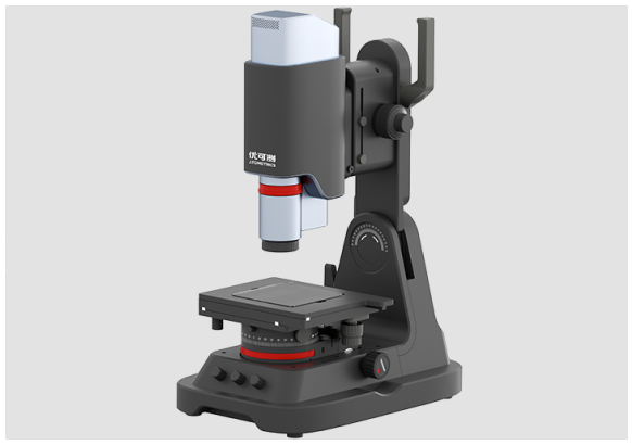
Atometrics Super Large Depth of Field Digital Microscope AH Series enables 360-degree, high-definition observation and analysis of surface defects, micro-topography, material metallographic structure, flaws/abnormalities, failure analysis, 2D/3D micro-measurement, etc. With a pixel count as high as 12 megapixels, it achieves one-click full-frame focused observation and measurement.

Boardstone lntelligent(Shenzhen)Co.,Ltd.
6F Building C Kaifa Plaza,7006 Caitian Rd.,Futian Dist,Shenzhen
Hotline:400 080 3885
Sales@atometrics.com.cn
WhatsApp: +86 13316897904